First #fluorescencefriday – the word of our expert
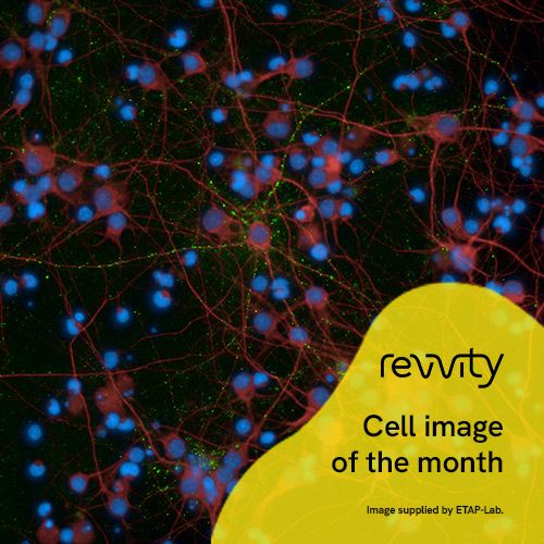
🔬 We wanted to share with you one of our cell image that shows primary cortical rat neurons (DIV14) stained for PSD-95 (synapses) in green, MAP2 (dendrites) in red and DAPI (nuclei) in blue. Each little green dot shows an active synapse happening between two neurons!
🧠Ahmad Allouche, one of our in vitro expert, explains:
“About the synaptic degeneration mechanism, it occurs early in neurodegenerative diseases and correlates with cognitive decline. Increased levels of synaptic proteins have been detected in the CSF in the early stages of Alzheimer’s diseases (AD) presumably due to release from degenerating synapses.
Postsynaptic density protein 95 (PSD-95, also known as SAP-90 or DLG4) is a scaffolding protein participating in the organization and regulation of synapses. It binds to NMDA and AMPA receptors, potassium channels, and associated signalling proteins resulting in their clustering during the formation and maintenance of dendritic spines. The dysregulation of PSD-95 expression has been associated with neurodegenerative disorders.
Reduced expression of PSD-95 has been observed in brain tissue from AD subjects and in mouse models of AD, making it an attractive target for developing strategies able to monitor PSD-95 accurately for therapeutics and diagnostics.“
Get in touch to develop your own study with our in vitro or in vivo models !
Image was acquired with Operetta® CLS™ High-Content Imaging System / REVVITY. Original picture © ETAP-Lab 2024.