Discover our platforms
Contact an expert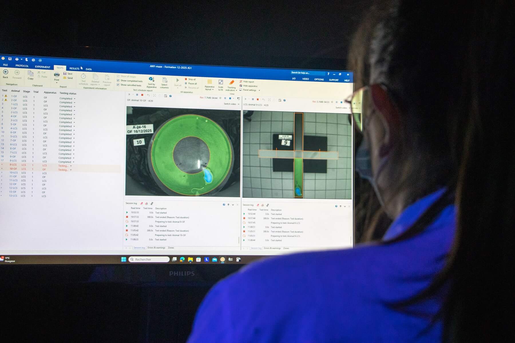
A single scientific partner delivering fully integrated, translation-driven solutions.
Our teams rely on cross‑functional technological platforms to provide you with seamless pharmacology services, from cell systems to large animals, ensuring continuity across every stage of your preclinical program.
- An end‑to‑end streamlined workflow: ETAP‑LAB fully internalizes immunohistology studies and multiplexed bioanalyses, giving you uncompromising control over data quality, assay reproducibility, and study timelines for model‑specific biomarker readouts.
- State‑of‑the art‑technologies with optimized cost efficiency: Access next‑generation in vitro high‑content screening and innovative microphysiological systems (MPS). Maximize the translational value of your drug candidate using high‑performance imaging modalities (MRI, PET, fUS, echocardiography) and clinically relevant behavioral endpoints.
- From cell systems to large-animal models: ETAP‑LAB operates under rigorously controlled laboratory conditions and within one of the most stringent regulatory frameworks worldwide for biosafety, biosecurity, and animal welfare.
OUR IN VITRO PHARMACOLOGY PLATFORMS
Molecule–Pathological Aggregate Interactions
This platform enables early-stage characterization of how your molecule interacts with the formation pathological protein aggregates involved in CNS proteinopathies such as Alzheimer’s or Parkinson’s disease.
Our differentiating strength lies in our proprietary know-how for the reproducible production and stabilization of human pathological oligomers (Aβ, α‑synuclein, Tau, TDP‑43). Each batch is qualified through QC procedures aligned with our internal standards, ensuring a reliable foundation for compound screening, whether for immunotherapies, small molecules, or other innovative approaches.
We routinely perform dot‑blot assays and protein aggregation kinetic studies (oligomer or fibril formations).
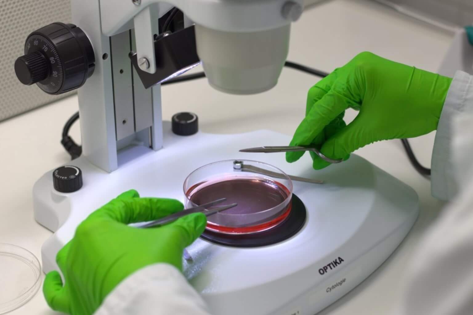
Cell Culture Laboratory
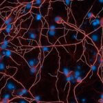
ETAP-LAB operates an L1 cell-culture facility dedicated to medium-throughput screening for neurology and stroke research. Direct adjacency to our animal facilities ensures quality and consistency of primary rodent neuronal cultures (mesencephalic, hippocampal, striatal, and cortical neurons).
ETAP-LAB is fully authorized to purchase, import, and store human‑derived biological materials (cell lines, iPSCs, samples…) enabling advanced cellular models.
Our facilities also allow the storage, re‑culturing, or preparation of your therapeutic innovations (explants, bioprinting constructs, reprogrammed cells, organoids…) prior to in vivo transplantation, supporting regenerative-medicine applications in Dermatology, Neurology, Neurovascular, and Cardiology.
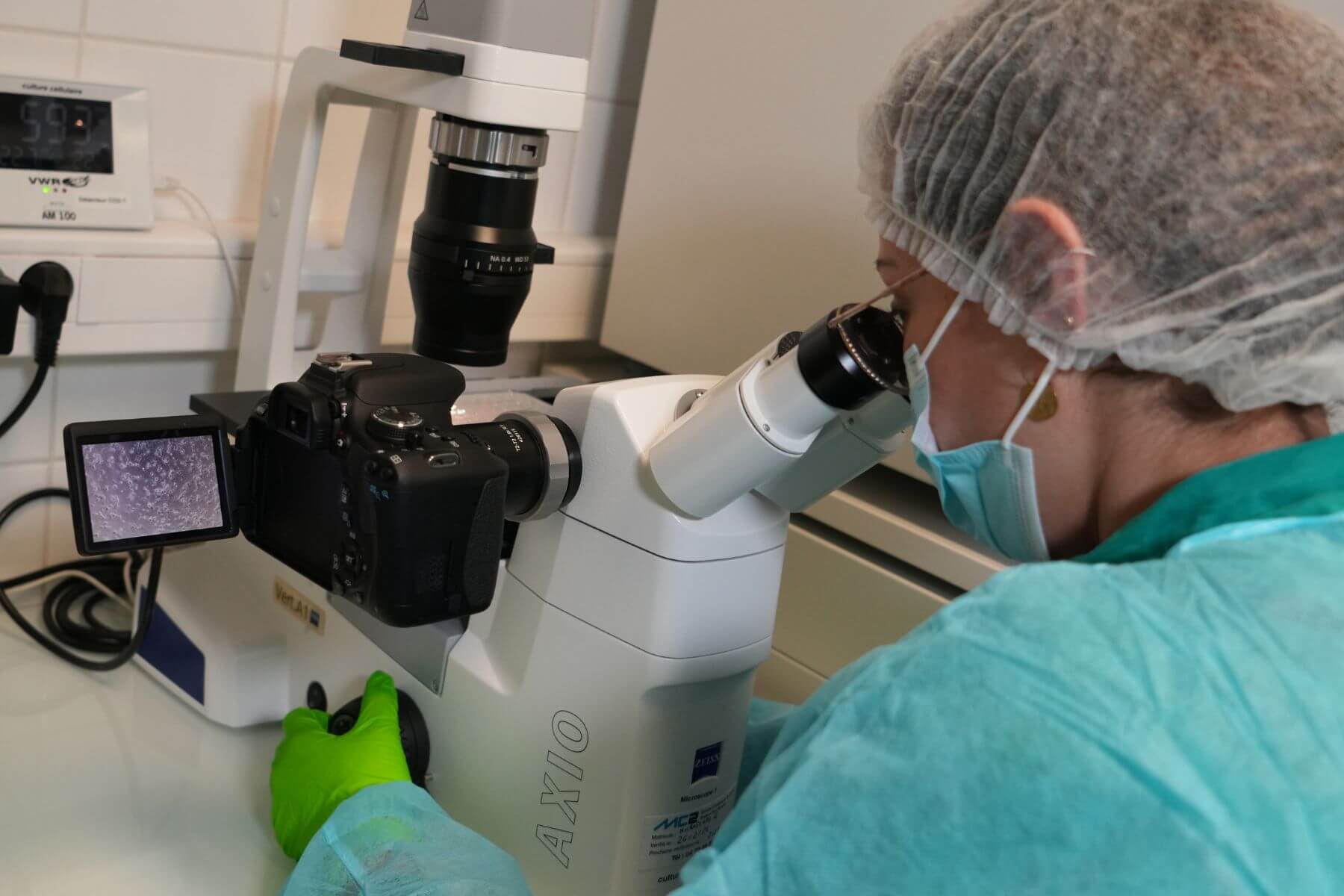
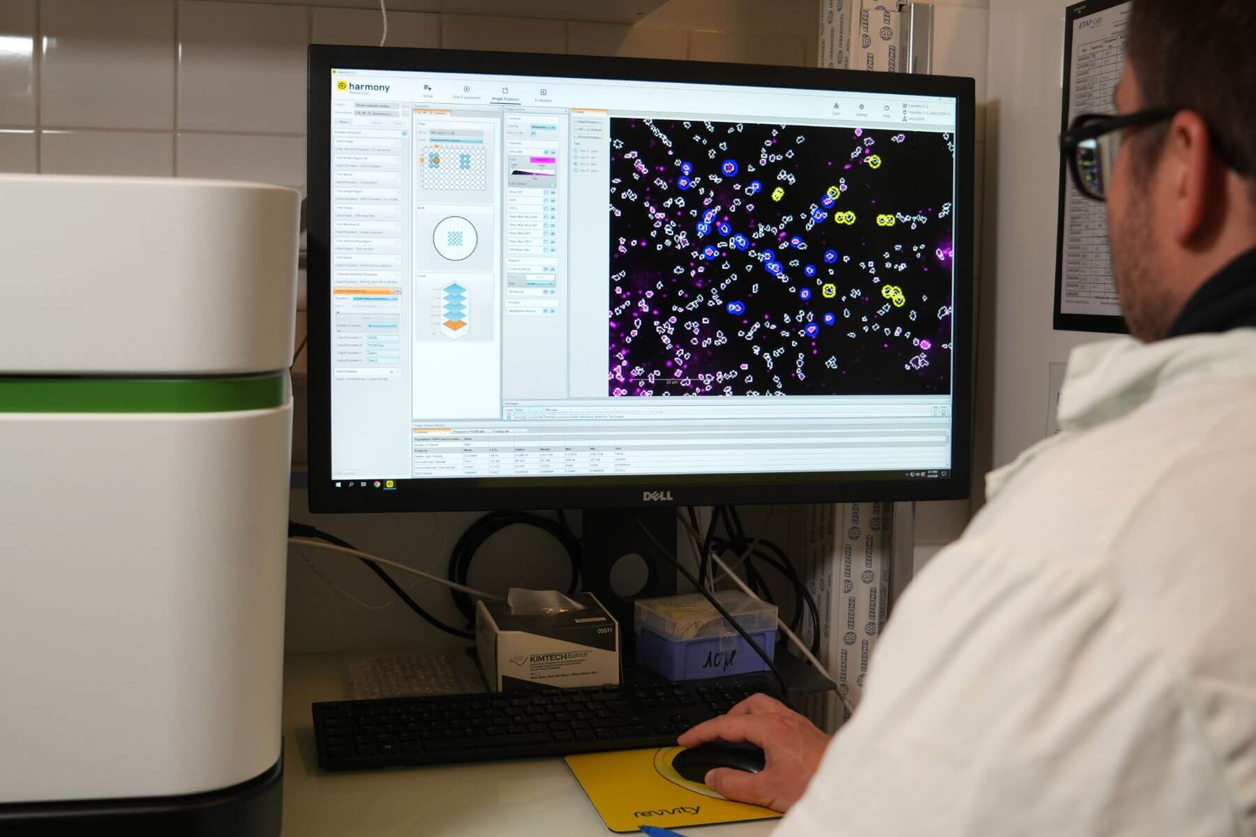
High-Content Imaging
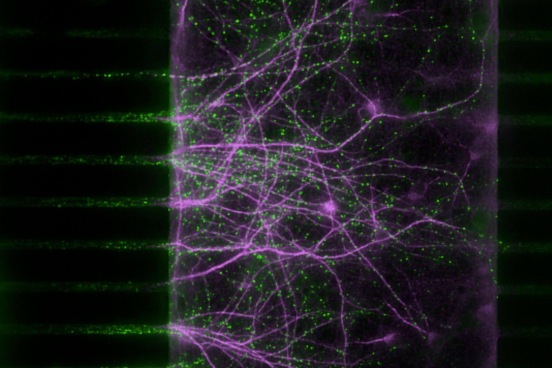
Using the Operetta CLS™ platform (Revvity), we automate medium‑throughput fluorescence imaging and integrate AI‑enhanced image‑analysis pipelines to deliver rapid, high‑accuracy quantification of large datasets.
Entrust your screening and lead-optimization campaigns to an expert team.
Brain‑on‑Chip platform
For several years, ETAP‑LAB has been investing in R&D programs aimed to engineer and scale-up brain‑on‑chip platforms, contributing to their routine adoption in preclinical CNS pharmacology.
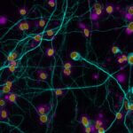
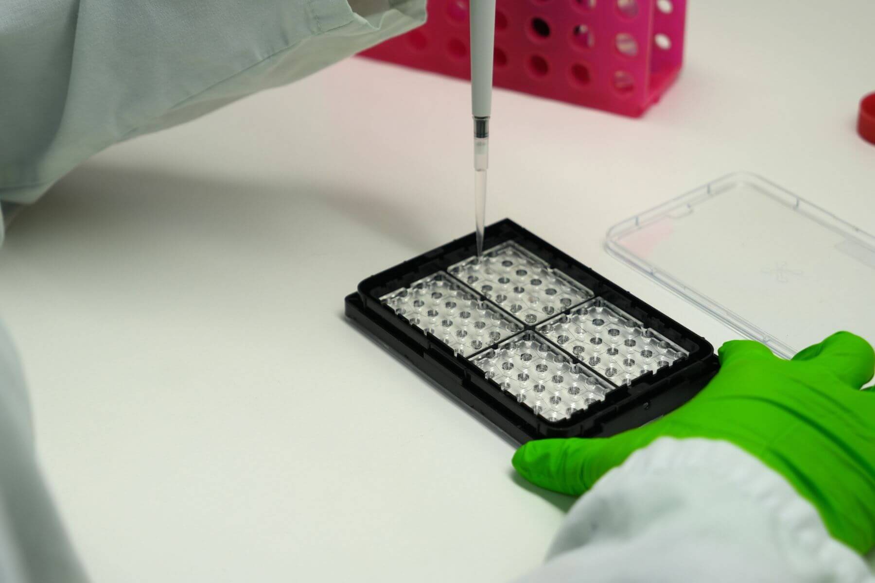
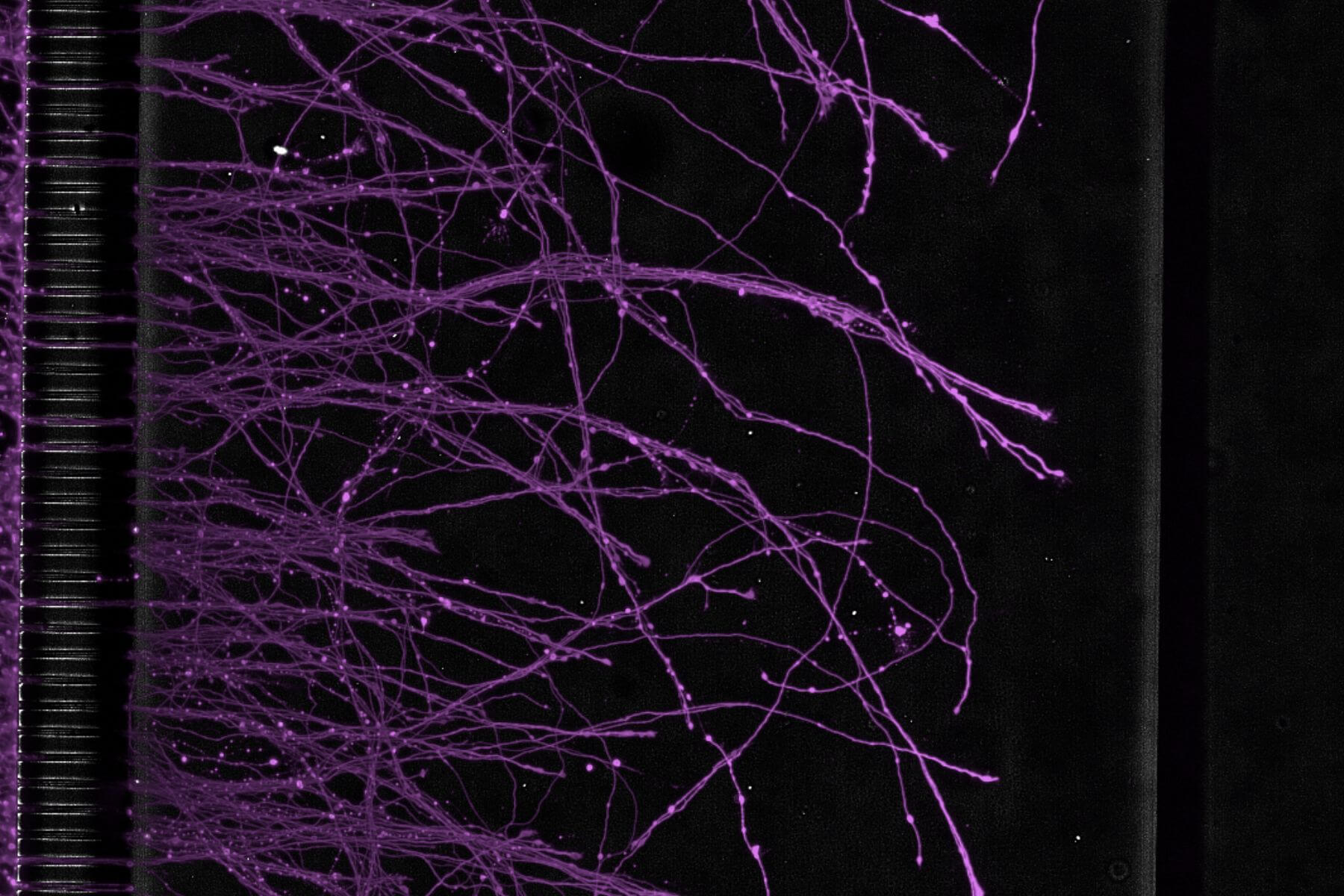
Built on compartmentalized microfluidic architectures, our brain‑on‑chip systems recreate the connectivity between spatially distinct neuronal populations maintained in different controlled microphysiological environments. This approach overcomes key limitations inherent to traditional 2D, 3D, and organoid cultures when modeling central nervous system biology.
In practice, our models enable targeted investigation of your drug-candidate effects on synaptic plasticity, axonal growth or degeneration, and axonal transport mechanisms – including pathological spreading – across interconnected neuronal populations.
All ETAP-LAB’s brain‑on‑chip models are compatible with High‑Content Imaging technologies and are available SBS‑formatted plates (16 chips per plate) ensuring reproducibility and a throughput suitable to pharmaceutical industry standards.
Brain-on-chips, and organ-on-chips more broadly, are microphysiological systems (MPS) recognized as new approach methodologies (NAMs) that support the current efforts to replace the use of animals for scientific purposes.
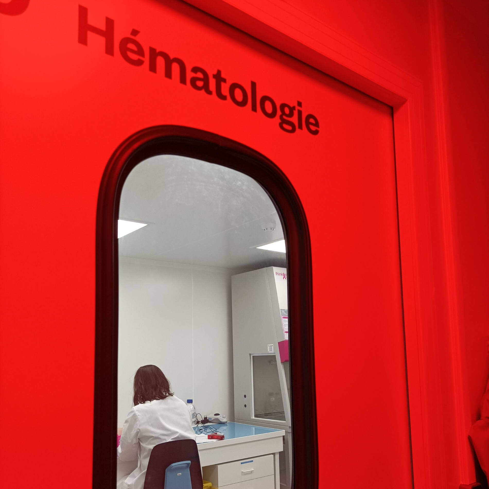
Hematology Laboratory
Our hematology laboratory is authorized to store and process human blood samples and derivatives for hemostasis and thrombolysis studies. ETAP‑LAB has an agreement with the French Blood Establishment (EFS), ensuring convenient local access to human blood samples for research applications.
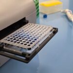
We offer whole‑blood ROTEM (rotational thromboelastometry) assays, as well as 96‑well turbidimetric assays using pooled human plasma. These tests deliver detailed data on coagulation dynamics and on the antithrombotic or fibrinolytic profiles of your drug candidates — either as standalone agents or in combination with reference standards.

OUR IN VIVO PHARMACOLOGY PLATFORMS
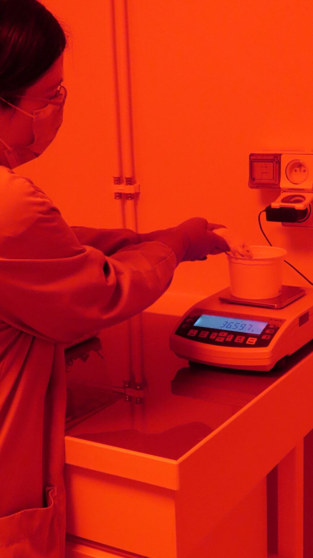
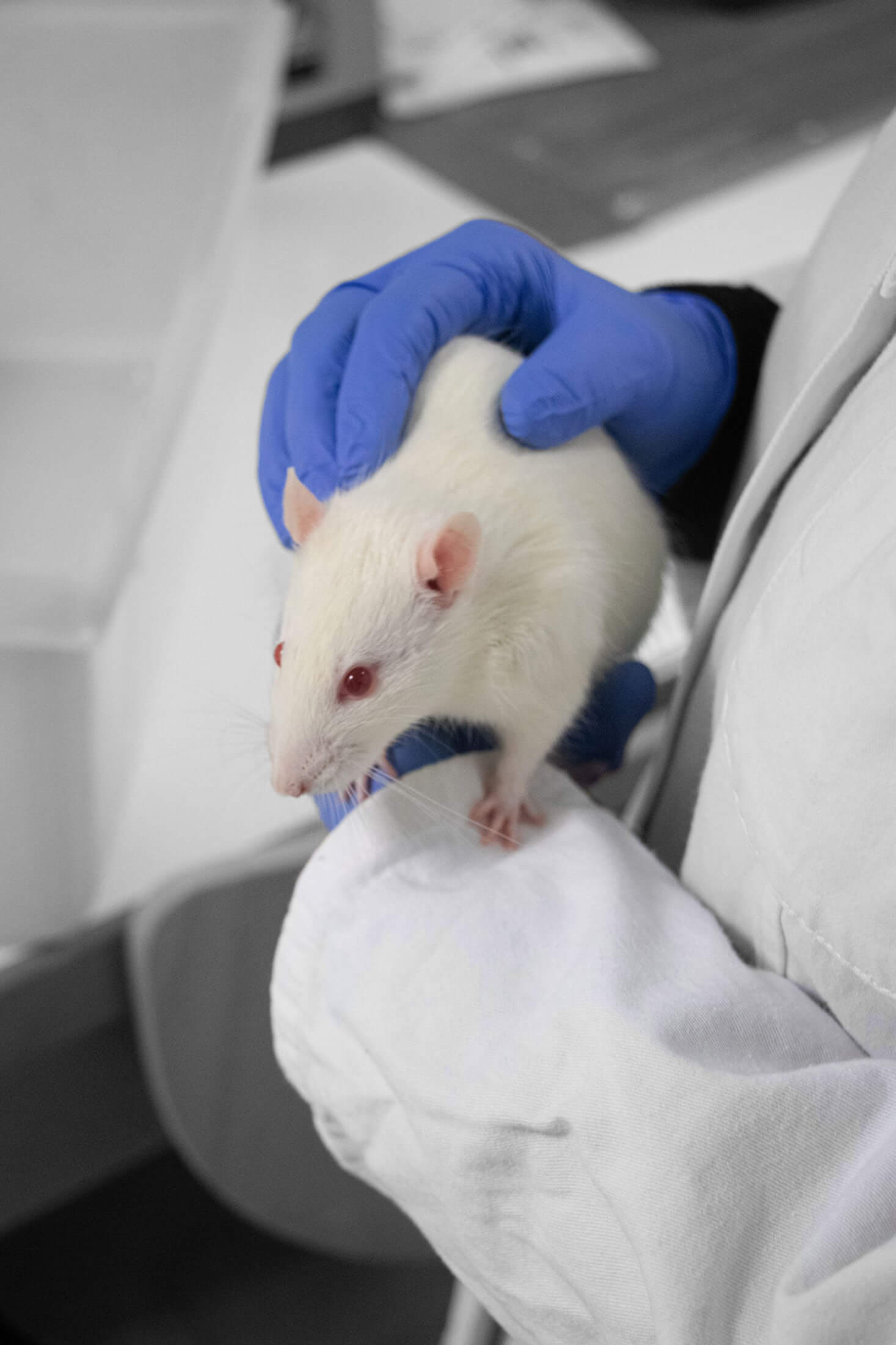
Small and Large Animal Facilities

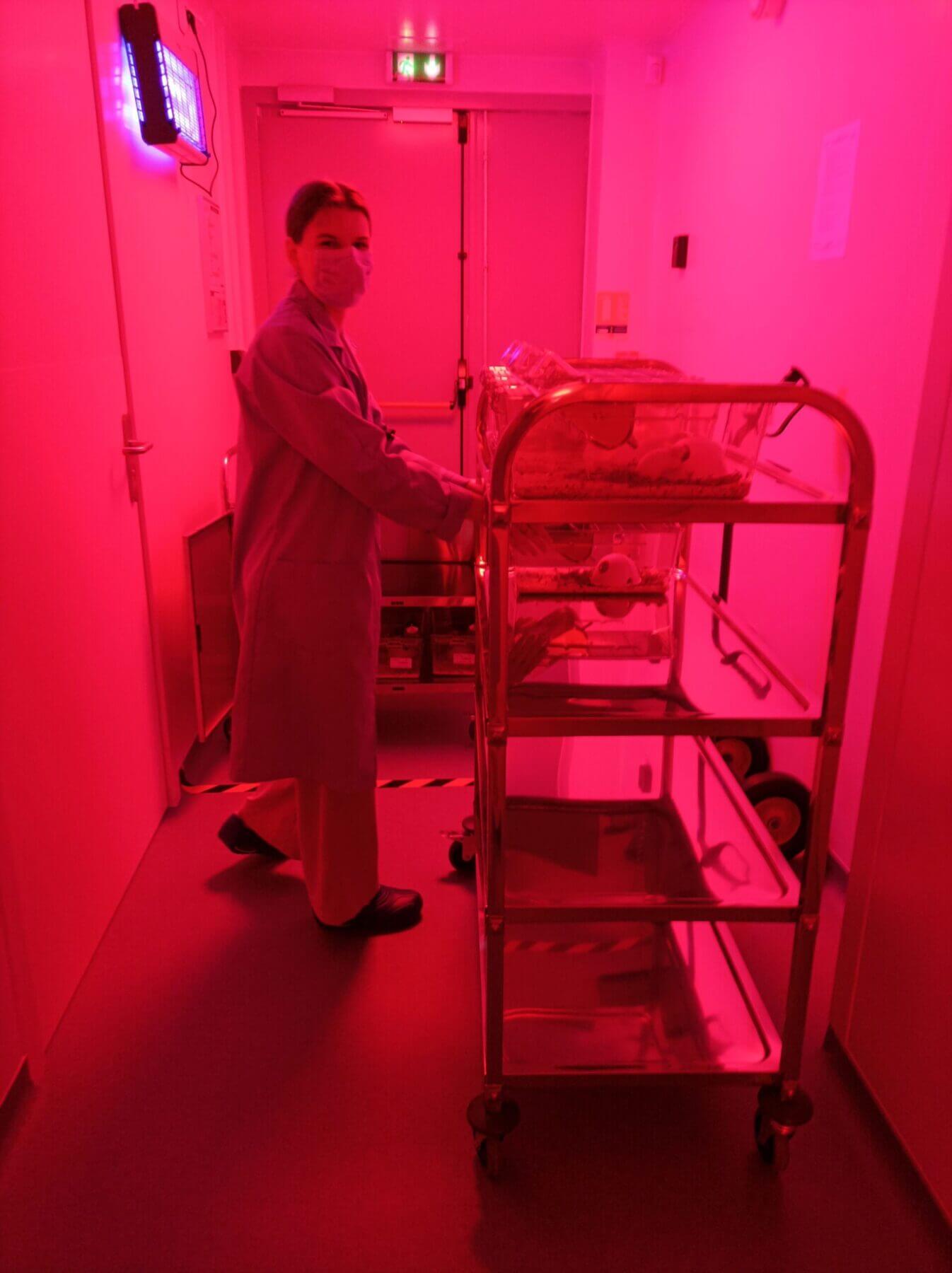
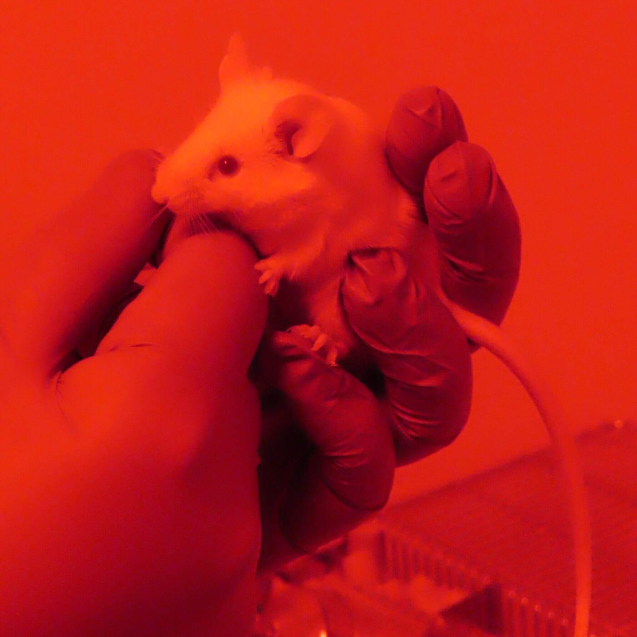
ETAP‑LAB operates 1,000 m² of fully accredited facilities (A1/A2 level), distributed across three sites with a total housing capacity of approximately 1,200 rodents. Our facilities are also authorized for Class 1 genetically modified mice and immunodeficient strains (NUDE and humanized).
Each facility includes controlled‑environment housing rooms, dedicated zootechnical areas, microsurgery platforms, and experimental rooms fully compliant with EU Directive 2010/63/EU.
All rodent studies are conducted under reversed light-dark cycle to align with rodent chronobiology and optimize the chronopharmacology of our preclinical trials.
We also have access to a large‑animal facility (rabbit, pig, goats, non‑human primates) equipped with both surgery and imaging platforms.
All studies obtained ethical approval by French Ministry of Higher Education and Research after detailed protocol review by an accredited animal research ethics committee and are conducted within one of the world’s most stringent regulatory frameworks for laboratory animal science. Discover our Ethics Commitments.
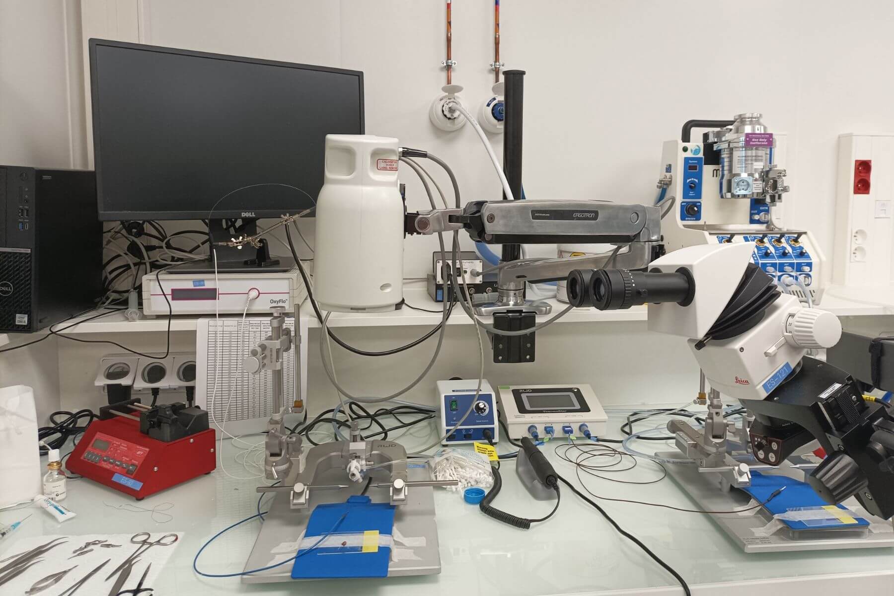
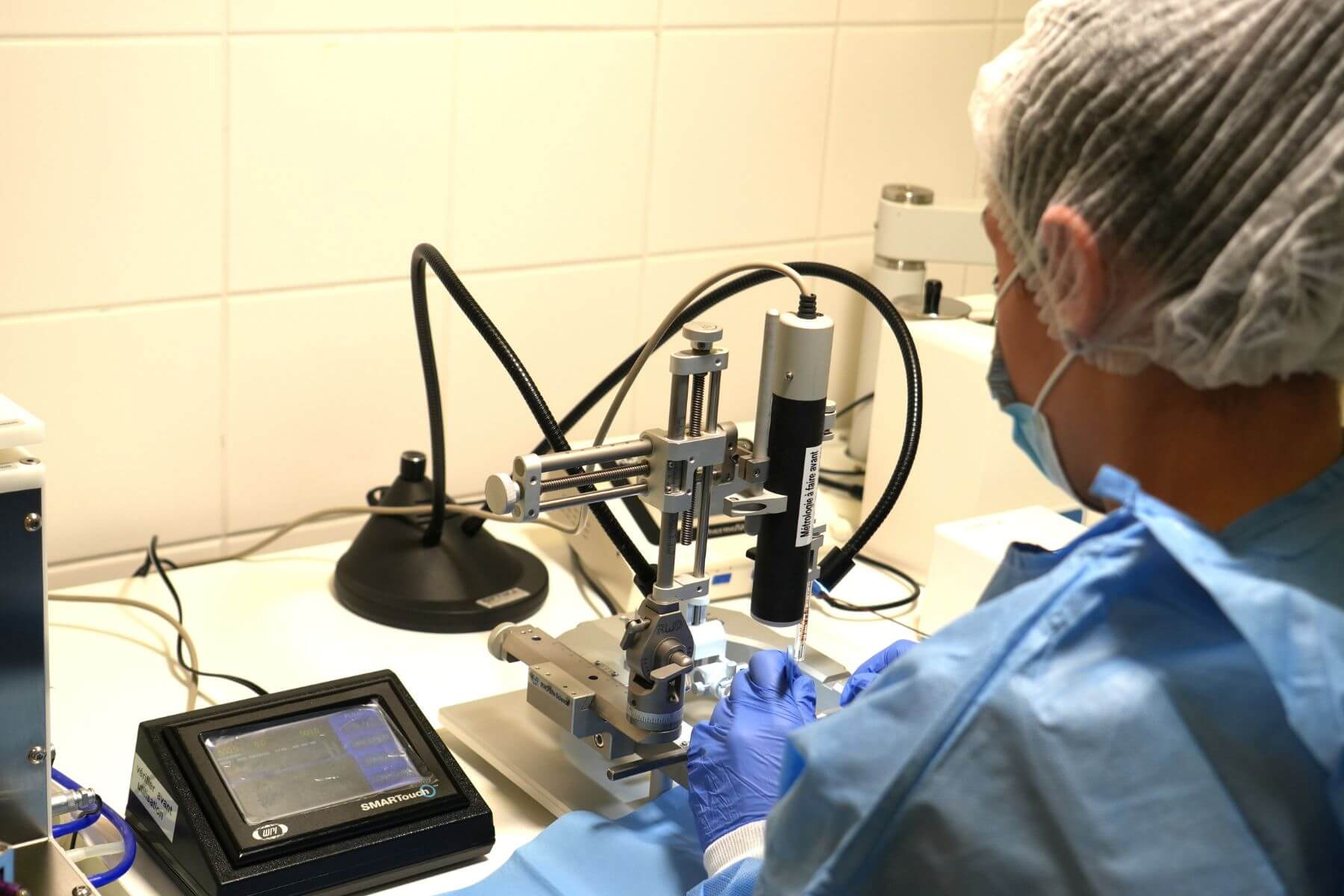
Stereotaxy and Microsurgery
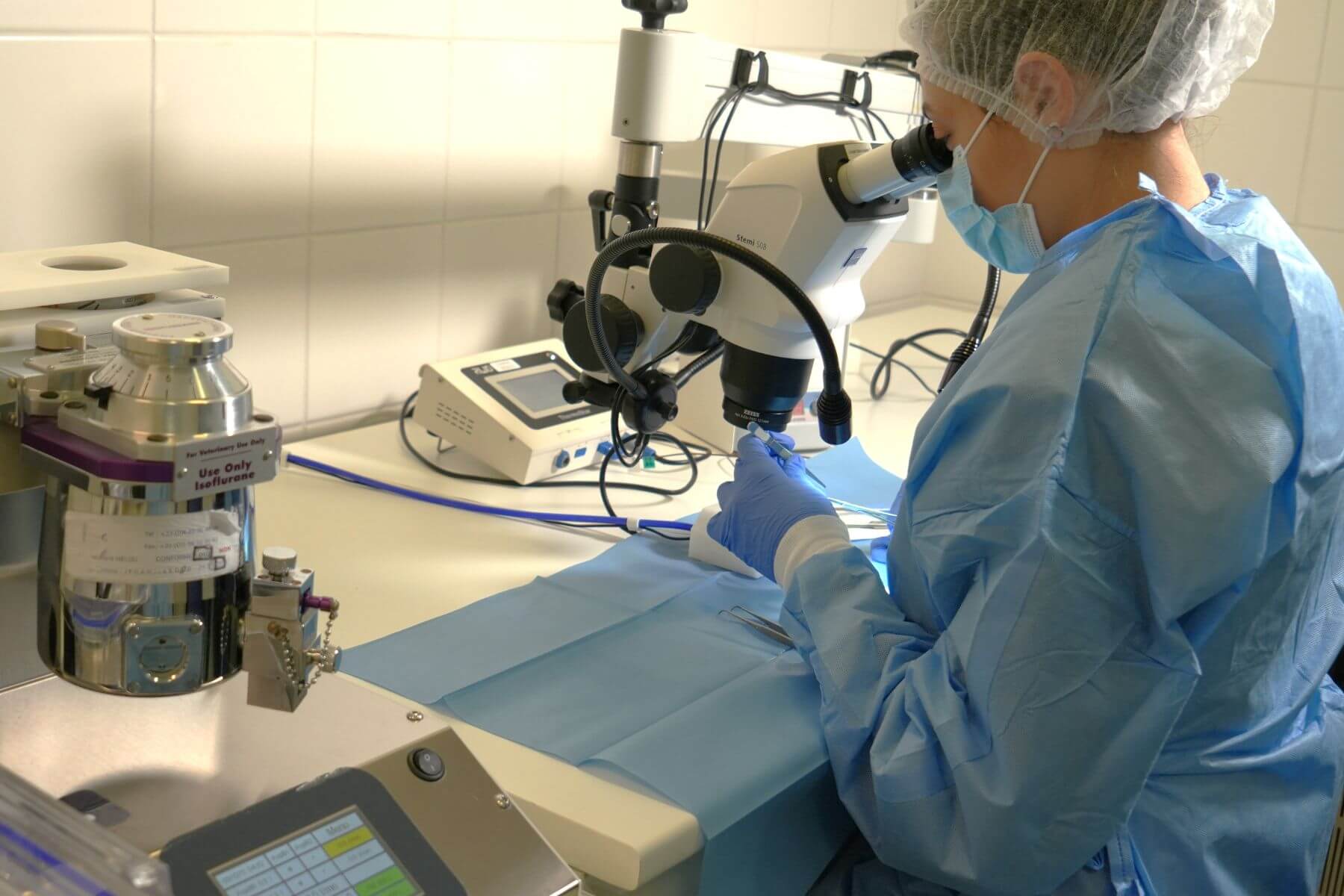
Our laboratories, audited by clients and veterinary referrals with excellent outcomes, include fully equipped operating rooms and specialized staff trained to rodent microsurgery. Procedures follow the highest standards across the entire perioperative workflow, including preparation, anesthesia, analgesia, asepsis, respiratory support, multimodal physiological monitoring, recovery, and post‑operative care.
Key equipment includes high‑magnification surgical microscopes for complex microsurgery (cardiac ischemia, skin grafting, MCA injection) and 12 stereotaxic stations enabling rapid deployment of Neurology protocols.
Beyond mandatory certifications, our surgeons complete an internal qualification program to minimize inter-operator variability in each surgical model inductions.
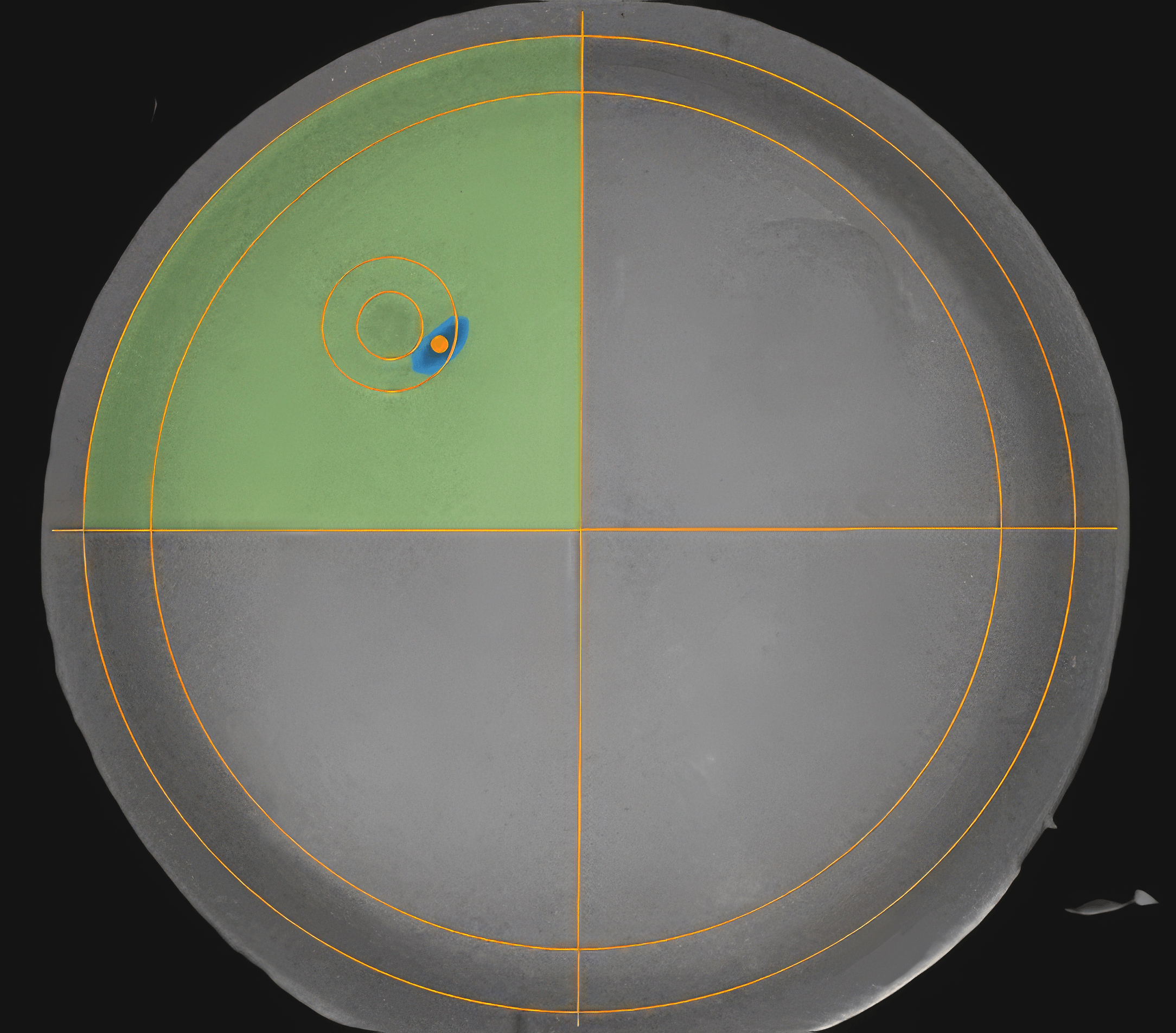
Behavioral Assessment Platforms
Our full range of behavioral tests in rodents allows the evaluation of motor, sensory, cognitive, mood‑related, motivational, preference‑based, well‑being, pain, and social interaction functions, and more. Learn more about our Ethological Best Practices.
These tests are validated as key readouts in our disease models in Neurology and Neurovascular research, as well as in Dermatology (e.g., itch) and Cardiology (e.g., incremental exercise test).
All platforms are designed according to best‑practice ethological standards: controlled chronobiology and light cycles, environmental enrichment, bias control (odor, noise, stress, lighting, handling), and precise definitions of behaviors.
Except when specific tests require otherwise, all behavioral sessions are video‑recorded and analyzed using automated video‑tracking for quick and objective analyses. Complex tasks (e.g., adhesive removal) are scored post hoc and blindly by trained staff. All videos are archived to ensure full traceability. Explore our Scientific publications.
OUR IN VIVO IMAGING PLATFORMS
Preclinical Medical Imaging
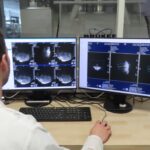
Through our long‑standing partnership with the CYCERON imaging platform in Caen, located directly next to our facilities, we have privileged access to state‑of‑the‑art medical imaging tools within an internationally recognized infrastructure. ETAP‑LAB teams – trained, autonomous, and experienced – routinely use the following systems:
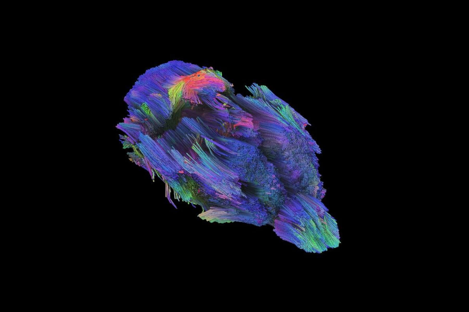
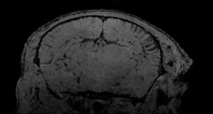
A 7T MRI for high‑resolution anatomical and functional imaging in rodents, with integrated physiological monitoring and motorized cradle for synchronized and reproducible acquisitions. Particularly valuable for Neurovascular studies, we offer numerous innovative sequences such as molecular imaging of inflammation.
A 3T MRI for large animals anatomical and functional imaging using a clinical-grade scanner.
A hybrid MRI‑PET system, combining 7T MRI spatial resolution with PET specificity, ideal for radiochemical biodistribution studies and small‑animal efficacy studies.
An MPI (Magnetic Particle Imaging), a cutting-edge whole‑body imaging technology with high temporal resolution for quantitative tracking of MPIO‑labeled molecules in PK or target-expression studies.
With more than 5,000 MRI procedures performed over the past five years, our teams demonstrate in-depth expertise in preclinical imaging acquisition, analysis, and interpretation.
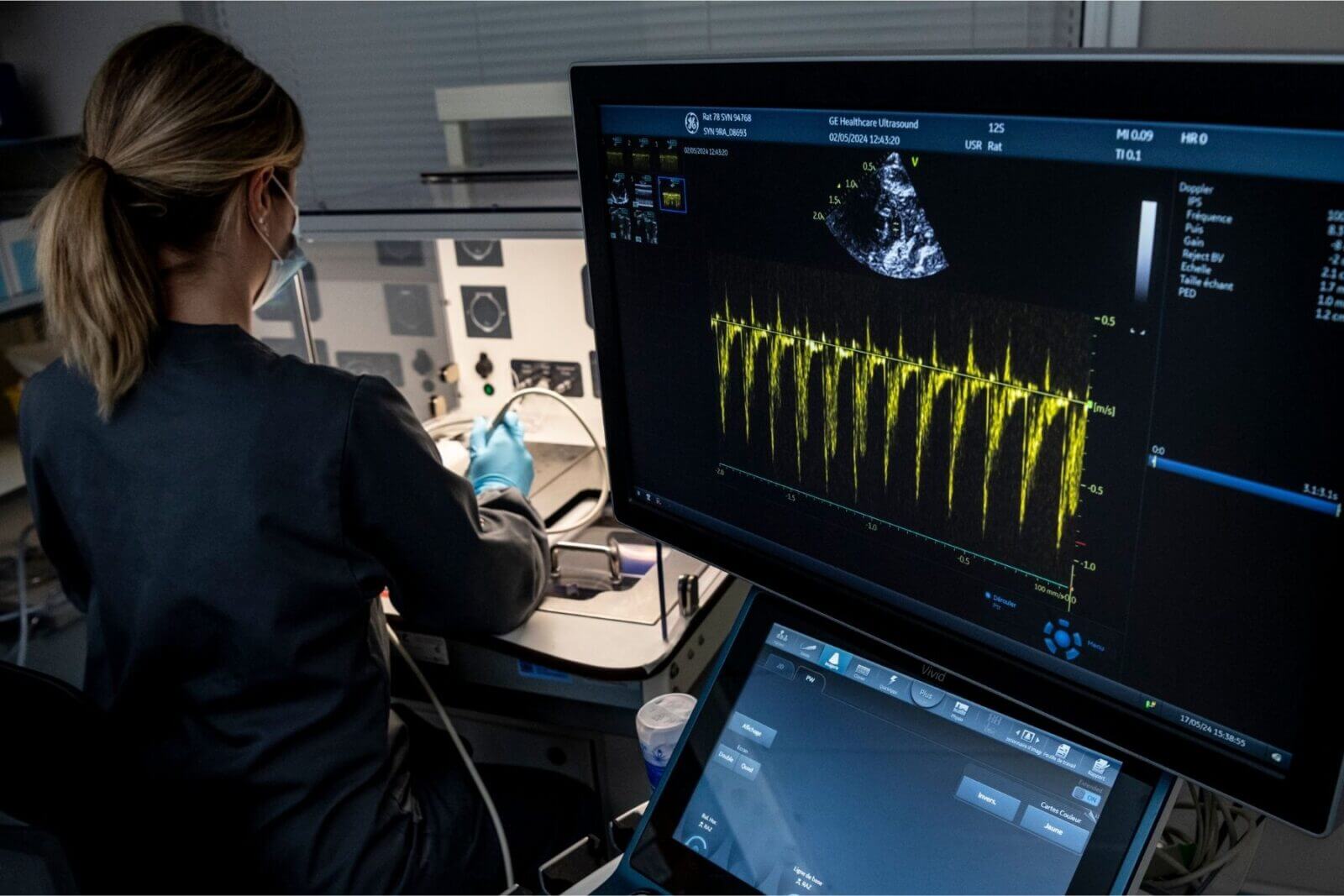
Ultrasound Imaging
We use two high-performances ultrasound systems with distinct applications:
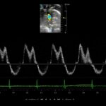
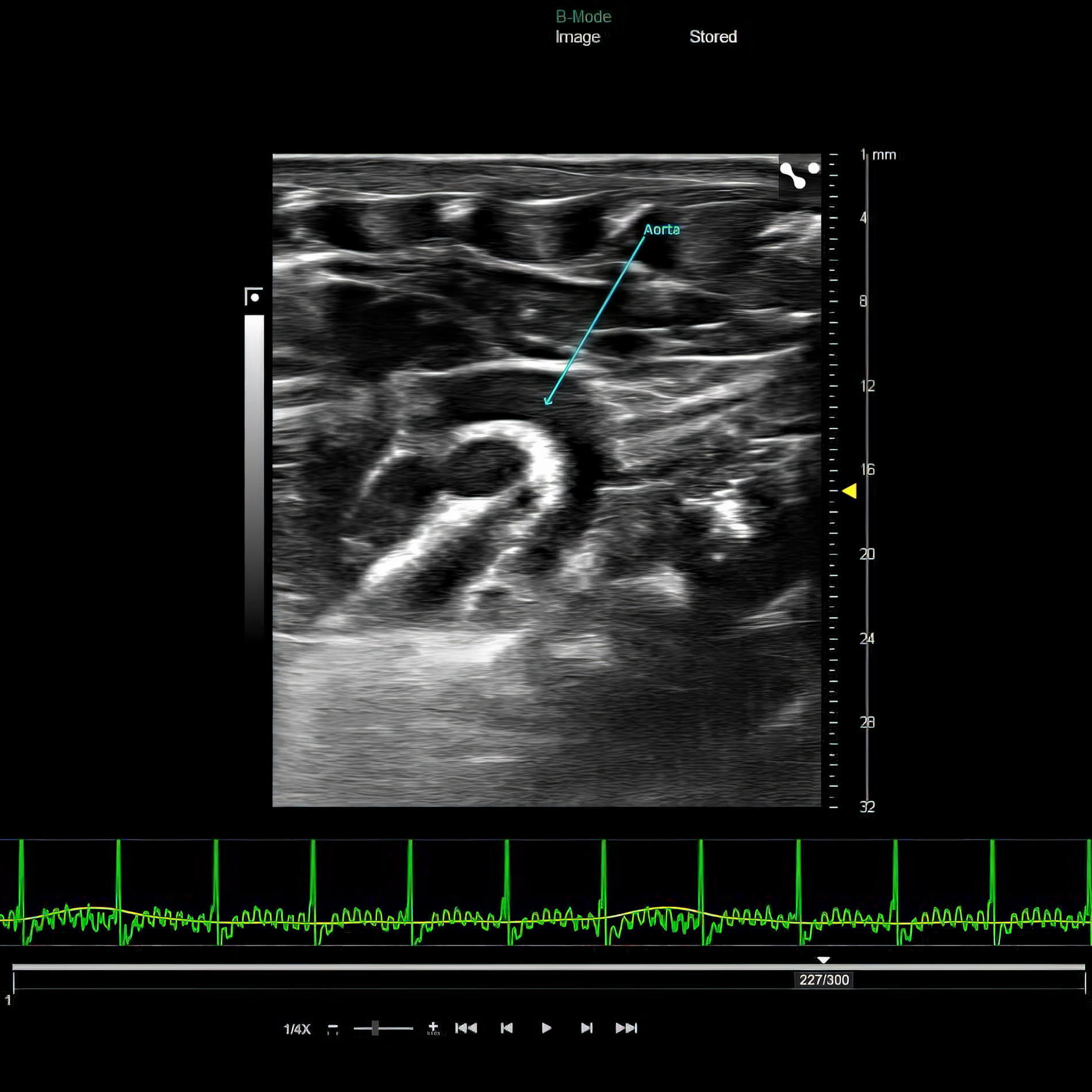
- A preclinical echocardiography system (VEVO) for preclinical cardiology,
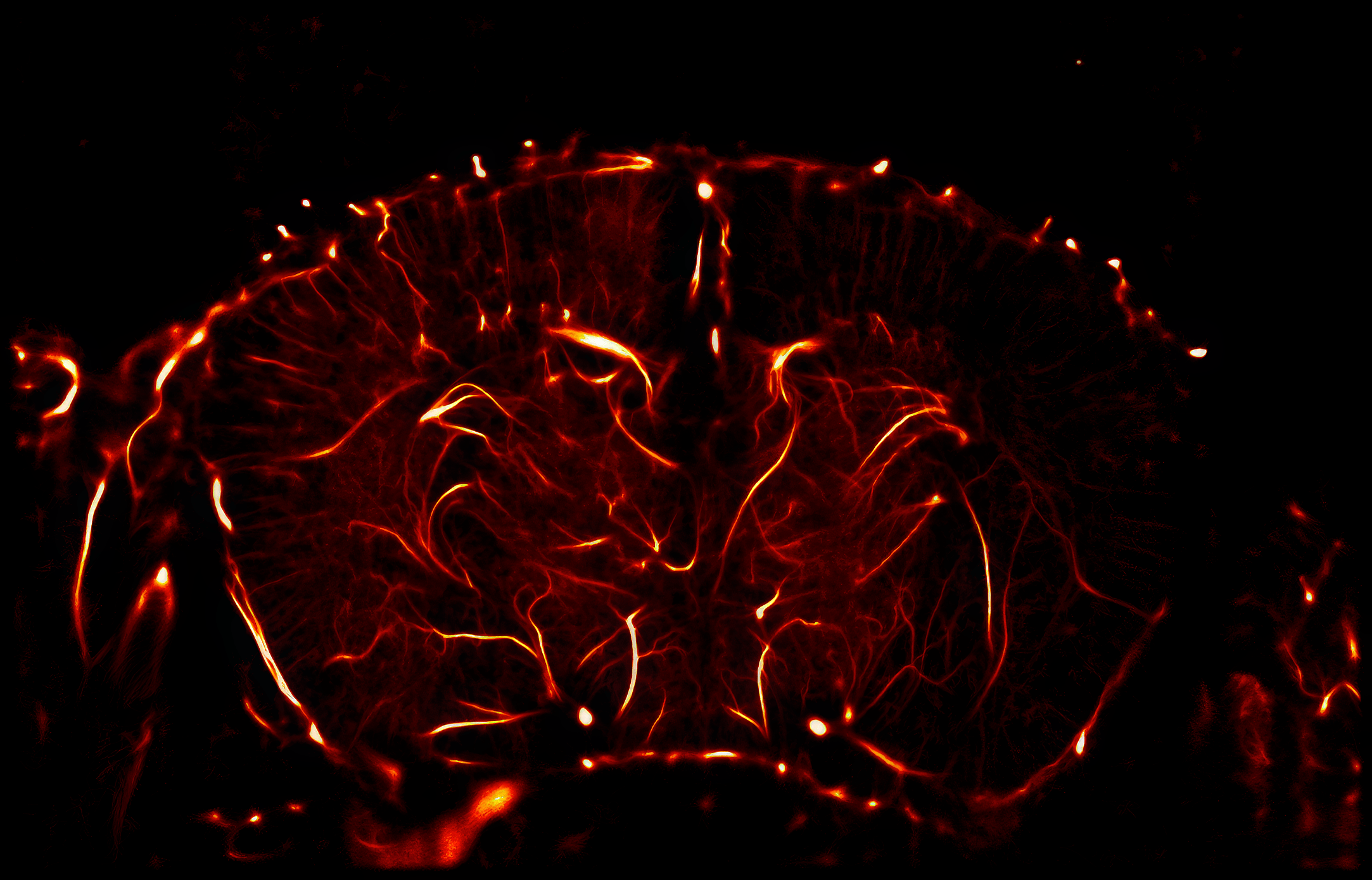
- A functional ultrasound (fUS) imaging system for neurovascular research.
The VEVO F2 echocardiography system is specifically designed for assessing cardiac function in small animals (down to Drosophila). Its ultra‑high‑frequency probes allow precise visualization of cardiac structures and their variations throughout the cardiac cycle in rats and mice. Its advanced Doppler modes provide detailed assessment of blood flow, pressure gradients, and regurgitations.
Functional ultrasound imaging enables dynamic mapping of brain activity with exceptional spatial and temporal resolution. This fully non‑invasive technique identifies localized variations in cerebral blood flow in response to external stimuli for functional imaging studies. Applications are numerous in neurovascular research: altered neurovascular coupling in pathological conditions, mapping of functional connectivity remodeling, visualization of cerebral perfusion including ischemic core and penumbra in stroke…
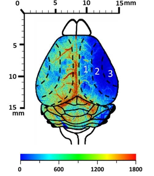
Laser Speckle Flow Imaging
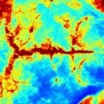
Laser Speckle Imaging delivers realtime high-resolution assessment of superficial tissue perfusion (depth ± 1 mm).
Particularly suited to our stroke models, this technology enables transcranial visualization of cerebral blood-flow variations over time across a large cortical area.
It also offers valuable applications in dermatology for monitoring skin-blood flow during wound healing, skin graft follow-up, or cutaneous ischemia events.
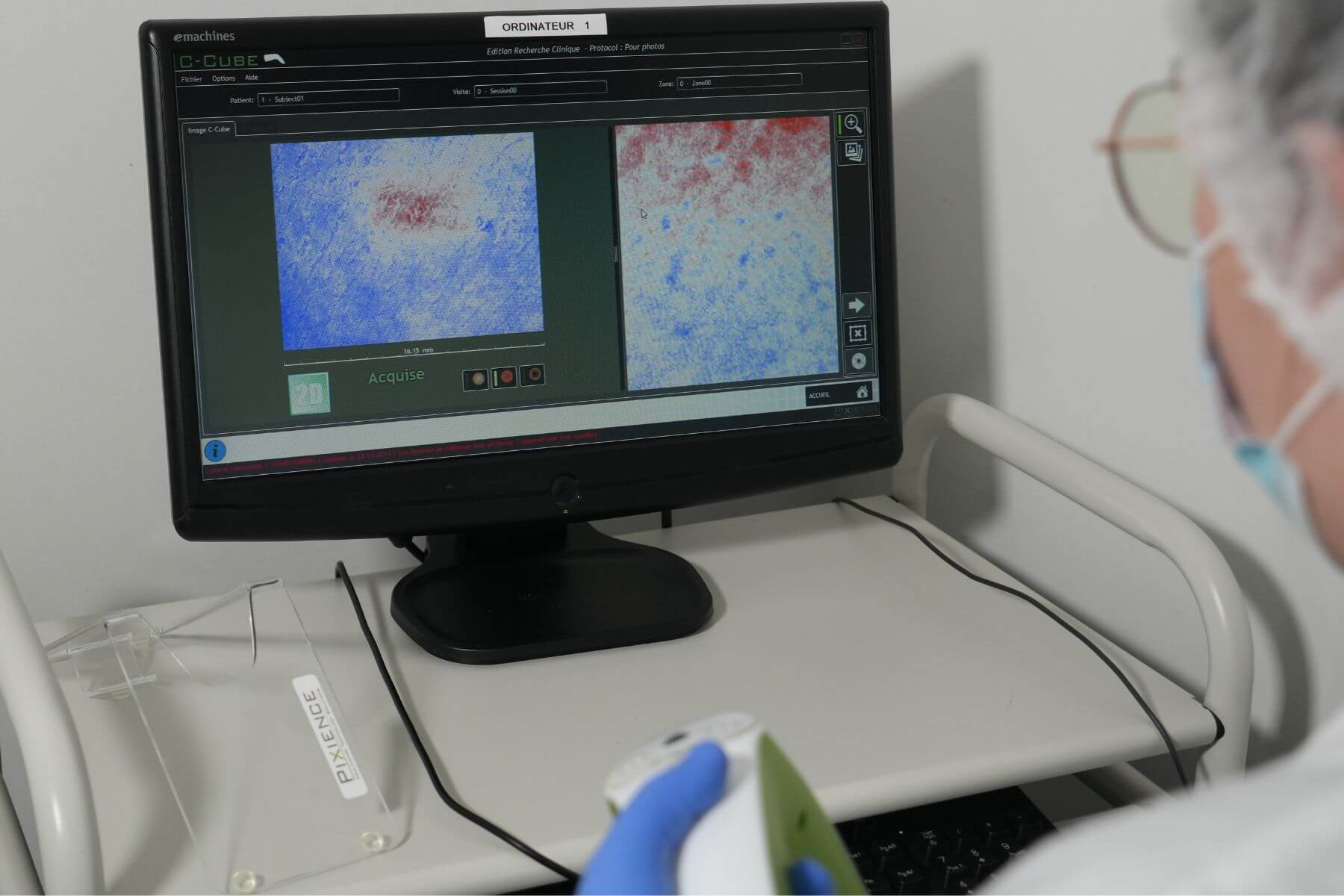
Dermoscopy
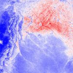
The C‑Cube dermoscope is a high‑resolution imaging system designed to capture the skin surface reproducibly in preclinical dermatology models.
Aligned with clinical standards, this technology enables precise documentation of texture, pigmentation, and lesion evolution over time. Its use strengthens translational value of your studies by providing reliable visual criteria comparable to clinical assessments. Integrated into our workflow, the C‑Cube dermoscope supports qualitative and quantitative evaluation of your therapeutic innovations in models of skin inflammation, skin barrier disruption, and skin grafts.
OUR BIOLOGICAL ANALYSIS PLATFORMS
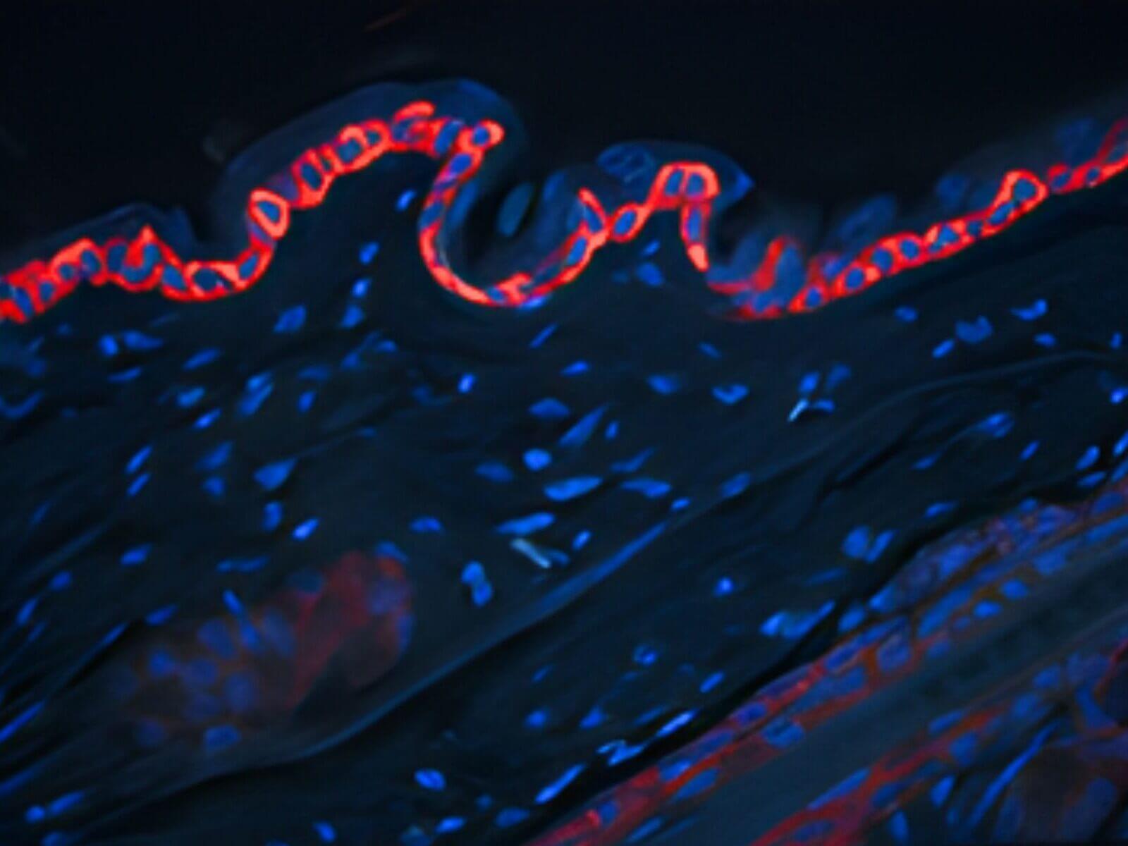
Immunohistochemistry, Immunofluorescence, and Histology
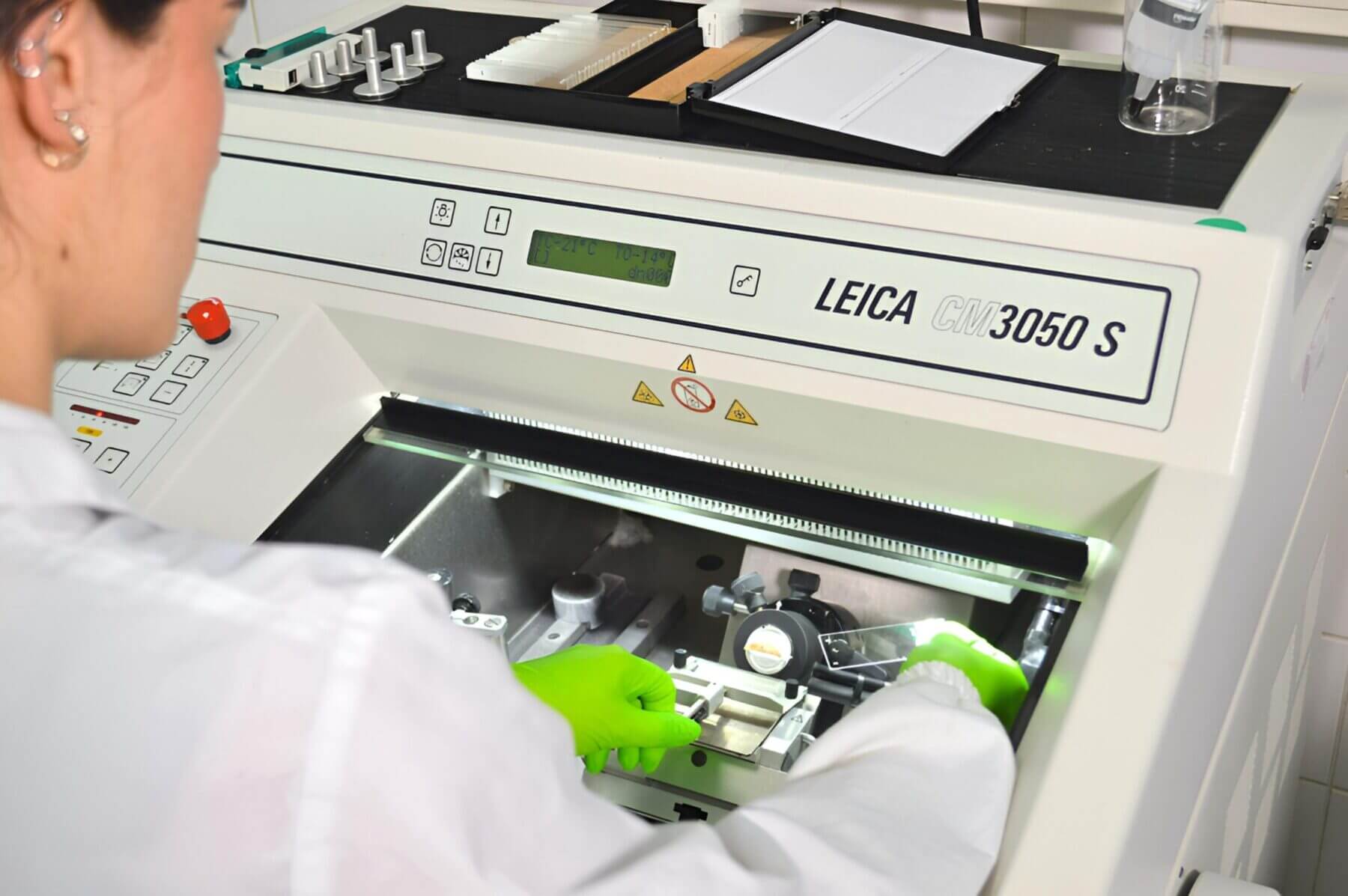
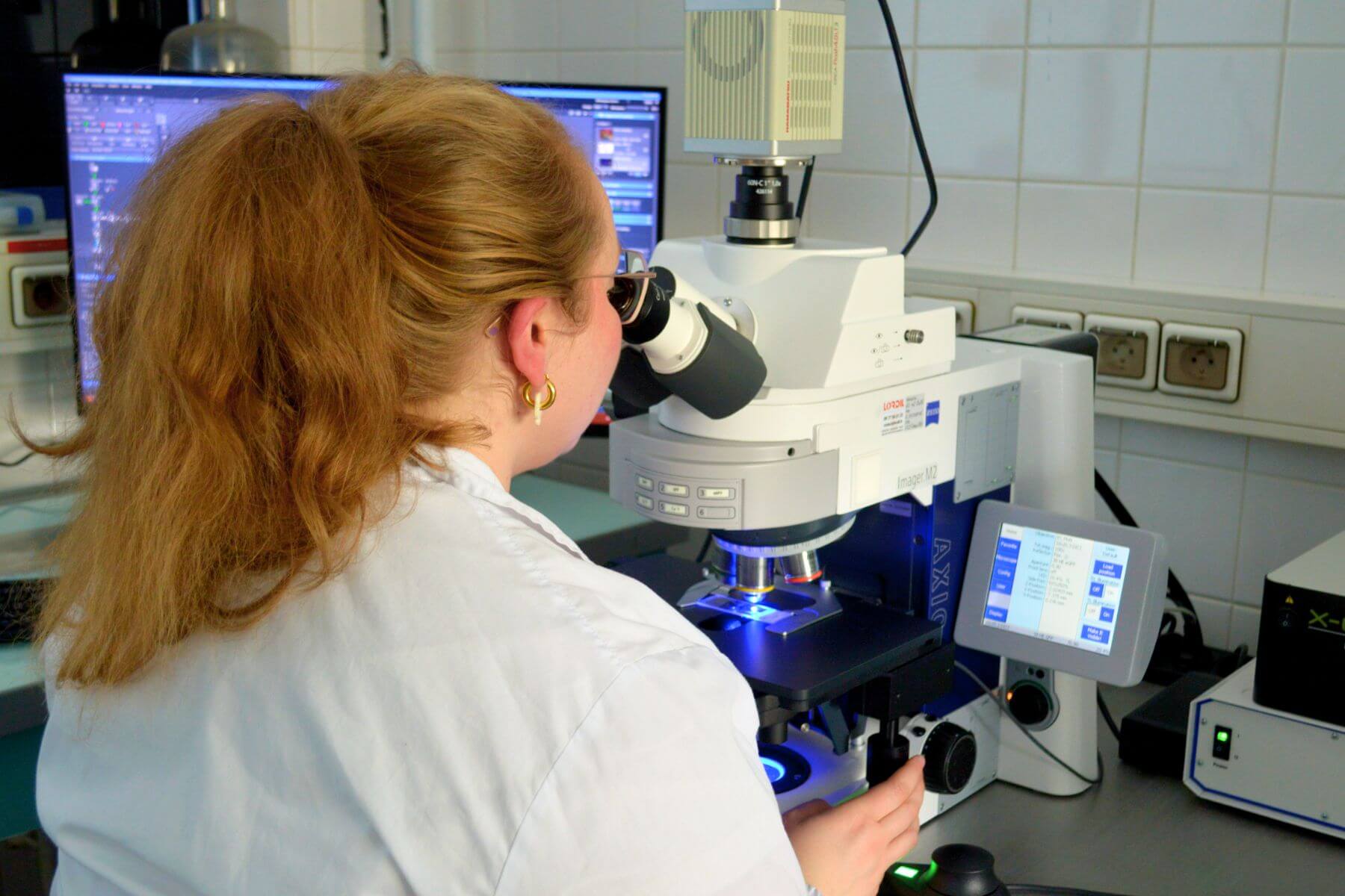
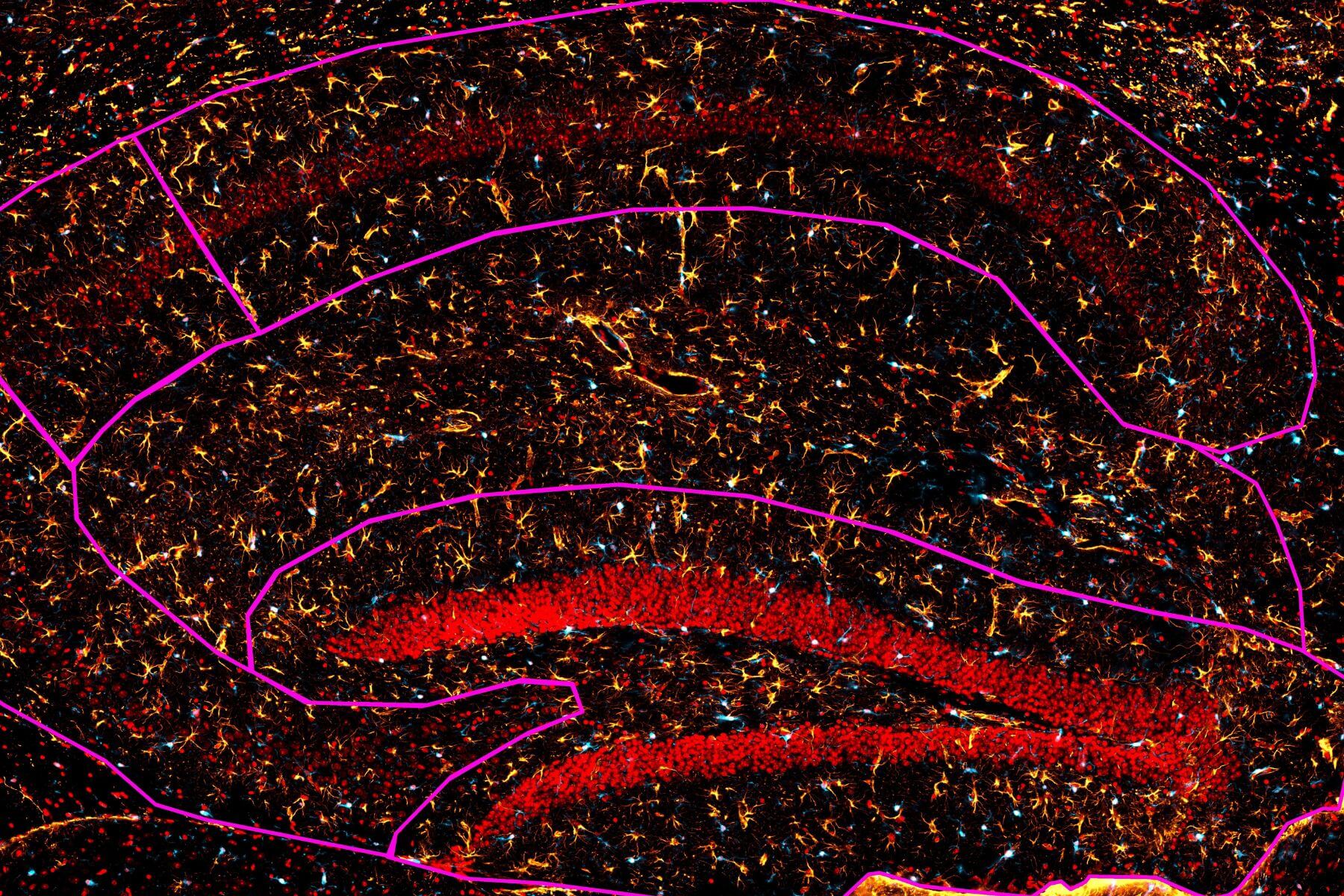
We process frozen tissues, including cryo‑embedding and customized cryo‑sectioning (thickness and orientation) using the Leica CM3050S. Image acquisition is performed in brightfield or multiplex fluorescence (up to 4 channels) using a Zeiss Axio Imager M2 automated microscope with z‑stack function for optimal data capture. Image analyses are performed semi‑automatically using dedicated software, while expert‑level slide review is available upon request for more complex or qualitative endpoints.
Our expertise spans a broad range of tissues including brain, heart, skin, liver, kidney, spleen, lung, and intestine.
Is your biomarker not yet available? We can develop a tailored staining and analysis workflow, including antibody validation and robust QC procedures.
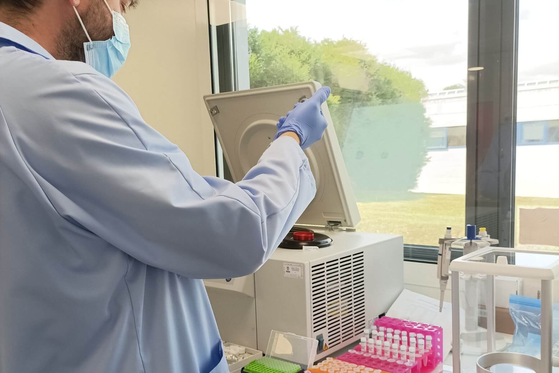
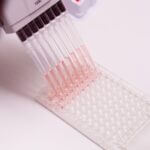
Immunoassays
Our integrated immunoassay platform ensures a continuous workflow coordinated with our in vitro and in vivo platforms, enabling uncompromised sample integrity, shortening timelines, and delivering reliable analyses across a panel of prevalidated biomarkers.
We routinely perform protein and enzymatic‑activity quantification using standardized colorimetric, fluorescent, and luminescent detection methods accross multiple sample types, including tissues, biological matrices, cell lysates, and culture supernatants.
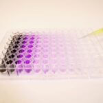
Our ELISA assays portfolio allows fast, quantitative approach to measure biomarkers variations associated with key pathological processes such as neurodegeneration, heart failure, immune activation, inflammation…
Our ELLA Simple Plex system (Bio‑Techne) provides high‑throughput, high‑sensitivity (< pg) multiplex quantification (up to 4 analytes) in minimal sample volumes (< µL), including a broad panel of mouse and human cytokines and chemokines.
Our capabilities also include routine Western blotting, including time‑resolved fluorescence (TRF) for ultra‑high‑sensitivity quantification in the femtogram range.
Is your biomarker not yet available? We can develop a tailored staining and analysis workflow, including antibody validation and robust QC procedures.
Additional Analyses and Outsourced Services

To extend our analytical capabilities, we rely on a network of trusted providers selected and regularly audited by our teams. Simplify the management of your studies with full peace of mind: we handle the relationship with the analytical provider on your behalf—from protocol definition to inclusion of results in our reports—and we commit to deliverable quality and timelines.
We collaborate routinely with veterinary and human pathology laboratories familiar with our preclinical models, ensuring quantitative and qualitative evaluation of biological endpoints.
We can also coordinate compound quantification by mass spectrometry for PK-PD and neuroPK studies. Our GLP Grade A partner develops and validates the analytical methods and performs the assays.
In addition, we can also coordinate RNA-Seq analyses (including single-cell), flow cytometry, and RNA immunohistochemistry by in situ hybridization.
We support you in your therapeutic projects and innovations.
ETAP-LAB is a preclinical CRO that provides efficacy testing and preclinical pharmacology services to the pharmaceutical, biotechnology, and nutraceutical industries.